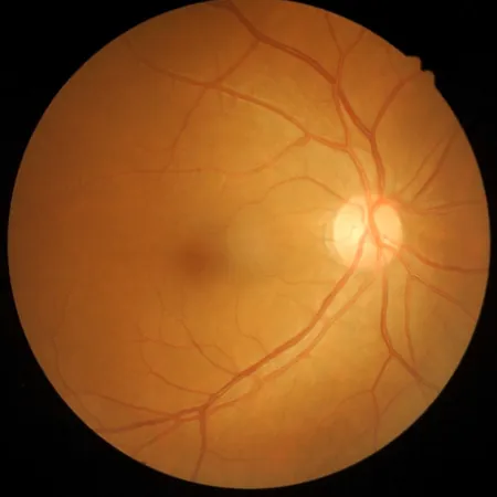
Study Reveals Alarming OCT Indicators for Retinal Atrophy Risk in AMD Patients
2025-05-25
Author: Michael
New Findings Could Change AMD Management Strategies
A groundbreaking study from Sapienza University of Rome has uncovered critical indicators that may signal an increased risk of retinal atrophy (RA) in patients with neovascular age-related macular degeneration (nAMD). Utilizing optical coherence tomography (OCT), researchers found that key features—such as type 2 macular neovascularization (MNV), thinning of the outer nuclear layer (ONL), reduced central foveal thickness (CFT), and the presence of intraretinal fluid (IRF) at baseline—hazily forecast a grim trajectory for these patients.
The Rising Tide of AMD
Age-related macular degeneration is the leading cause of blindness in many industrialized nations, with projections estimating an increase from 196 million cases in 2020 to a staggering 288 million by 2040. The complex nature of AMD varies by geographical location, warranting deeper understanding beyond demographic differences.
Predicting Progression: A Must for Effective Treatment
Dr. Oscar M. Gagliardi and his team have emphasized the urgent need for reliable markers to gauge AMD progression, which can drastically improve treatment plans and preventative strategies. In their study, they meticulously analyzed OCT characteristics that may predict RA in patients receiving aflibercept treatment.
In-Depth Study Insights
This retrospective observational study examined 1,085 clinical records, narrowing down to 703 patients who were treated with aflibercept. With rigorous follow-up, the analysis included 46 eligible eyes, averaging 78.37 years in age. Remarkably, while some eyes showed no signs of RA after five years, 25 eyes met the established criteria for RA.
Dramatic Visual Outcomes at Five-Year Mark
While no significant age difference was recorded between the groups, visual acuity outcomes told a dramatic story. At the five-year mark, patients with atrophy suffered a significantly worse best-corrected visual acuity (BCVA), highlighting the dire consequences of RA.
Diving Deeper: OCT Findings
The study's findings revealed that patients in group B (those with RA) exhibited a thinner ONL and reduced CFT—critical indicators that serve as alarming warnings for ophthalmologists regarding the disease's progression. While both groups had similar MNV characteristics, the prevalence of type 2 MNV was notably higher among atrophic eyes.
Future Research Directions
Despite the promising results, Gagliardi's team pointed out potential limitations, such as the reliance solely on OCT imaging for defining RA. They noted the importance of exploring additional imaging technologies and treatment options to create a more robust understanding of AMD.
Conclusion: A Call for Comprehensive Approaches
With AMD cases on the rise, these findings underscore an urgent call to action for ophthalmic researchers and clinicians alike. The integration of multiple imaging modalities and evaluation of various anti-VEGF treatments could pave the way for more effective management strategies, ultimately improving outcomes for millions at risk.


 Brasil (PT)
 Canada (EN)
 Chile (ES)
 Česko (CS)
 대한민국 (KO)
 España (ES)
 France (FR)
 Hong Kong (EN)
 Italia (IT)
 日本 (JA)
 Magyarország (HU)
 Norge (NO)
 Polska (PL)
 Schweiz (DE)
 Singapore (EN)
 Sverige (SV)
 Suomi (FI)
 Türkiye (TR)
 الإمارات العربية المتحدة (AR)