Revolutionary Surgical Microscope Uses 48 Cameras for Unmatched 3D Imaging
2025-07-07
Author: Noah
A Game-Changer for Surgeons
For over a century, surgeons performing intricate procedures have relied on traditional stereoscopic microscopes to provide a sense of depth. These devices, mimicking human vision, have allowed doctors to perceive three-dimensional structures essential for navigating complex bodily landscapes—especially delicate blood vessels and intricate brain tissues.
While advancements such as digital displays and video functionality have modernized these microscopes, the fundamental principle remains unchanged: two views interpreted by the human brain.
The Limitations of Conventional Imaging
However, this traditional approach comes with significant constraints. Although it offers decent depth perception, extracting precise measurements from two images—especially in chaotic surgical environments—is anything but straightforward. Factors like uneven lighting, reflective surfaces, and obstructive tools can complicate procedures, limiting the effectiveness of surgical automation and real-time feedback tools.
While preoperative 3D scans such as MRIs or CTs provide some guidance, they lack real-time updates as tissues shift during surgery. Optical coherence tomography (OCT) offers detailed insight but only for small areas and often produces challenging black-and-white images.
Introducing the FiLM-Scope: A Breakthrough in Surgical Imaging
In response to these challenges, researchers have unveiled the "FiLM-Scope," a pioneering surgical microscope that harnesses the power of 48 tiny cameras. Recently highlighted in Advanced Photonics Nexus, this innovative device is a potential game-changer for both surgeons and robotic instruments.
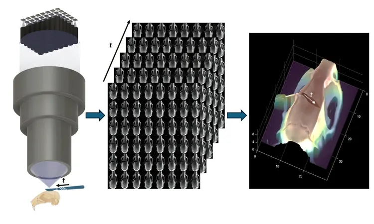
The FiLM-Scope employs a grid of 48 cameras, all focused through a single, high-performance lens. These cameras capture the surgical scene from slightly different angles, generating an impressive 48 high-resolution images—each boasting 12.5 megapixels. With a wide field of view of about 28 by 37 millimeters and intricate details down to 22 microns, it also streams video at a rapid 120 frames per second.
Real-Time 3D Mapping at Your Fingertips
The advanced imaging solutions of FiLM-Scope don't stop there. A specially designed self-supervised algorithm processes these multiple perspectives to create a precise 3D map in real time. This means it can reconstruct surface shapes with astounding accuracy—up to 11 microns over a depth range of one centimeter.
What sets FiLM-Scope apart is its ability to allow users to digitally zoom or shift the view without moving the microscope itself. This innovation promises to enhance the surgical experience, making procedures smoother and more efficient.
Broadening Horizons in Microsurgery and Beyond
Transforming standard images into exact 3D measurements, the FiLM-Scope holds the potential to elevate both manual and robotic microsurgery to unprecedented levels. Its versatile and data-rich imaging capabilities could also find applications across diverse fields that require high-precision 3D visualization—from materials science to microfabrication. The future of surgery may just be here, and it looks more precise than ever!


 Brasil (PT)
 Canada (EN)
 Chile (ES)
 Česko (CS)
 대한민국 (KO)
 España (ES)
 France (FR)
 Hong Kong (EN)
 Italia (IT)
 日本 (JA)
 Magyarország (HU)
 Norge (NO)
 Polska (PL)
 Schweiz (DE)
 Singapore (EN)
 Sverige (SV)
 Suomi (FI)
 Türkiye (TR)
 الإمارات العربية المتحدة (AR)