Groundbreaking Discovery: Scientists Capture 3D Footage of Heart Formation in Mice Like Never Before!
2025-05-13
Author: Jacques
Revolutionary 3D Imaging Unveils Heart Development
In an astounding breakthrough, researchers from UCL and the Francis Crick Institute have captured 3D images of a heart forming inside a living mouse embryo, revealing the origins of cardiac cells like never before.
Advanced Techniques Pave the Way for Major Discoveries
Published in *The EMBO Journal*, this groundbreaking study employed advanced light-sheet microscopy on a specially engineered mouse model. This innovative method uses thin sheets of light to illuminate tiny samples, allowing scientists to create detailed 3D images without harming living tissue.
Tracking Heart Cells in Real-Time
Researchers were able to track individual cells as they moved and divided over 48 hours, from the pivotal stage of gastrulation, where cells begin to organize into vital structures, to when the primitive heart starts forming. This crucial insight opens new avenues for understanding the cellular origins of the heart.
The Importance of Gastrulation
Gastrulation occurs in humans during the second week of pregnancy and is essential for establishing the body’s primary structures, including the heart. The findings hold great promise for revolutionizing how scientists approach congenital heart defects, which affect nearly one in 100 babies.
Unexpected Findings and New Understanding
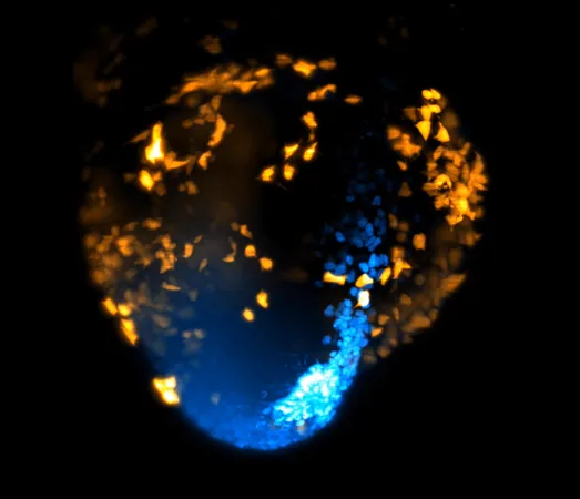
Dr. Kenzo Ivanovitch, senior author of the study, stated, 'This is the first time we've been able to watch heart cells this closely, for this long, during mammalian development.' By using fluorescent markers, the team tagged heart muscle cells, allowing them to produce a detailed time-lapse video that captured every movement and transformation.
Mapping the Cell Family Tree
Snapshots were taken every two minutes, producing unprecedented images of how cells coalesce to form the heart. This created a ‘family tree’ of cells, helping the researchers identify when and where the first heart-specific cells appeared in the embryo.
Controlled Chaos: A New Perspective on Cell Movement
The study revealed that embryonic cells are multipotent at first, capable of becoming various types, including heart and blood vessel cells. However, only hours into gastrulation, cells destined for the heart emerge with surprising organization, following specific paths rather than moving randomly. Dr. Ivanovitch explained that this shows cardiac development is governed by underlying patterns.
Future Implications for Medicine
Lead author Shayma Abukar described the team's efforts to decode the signals orchestrating these complex cell movements. She emphasized that the heart arises from various distinct cell groups appearing at different times during gastrulation, fundamentally changing our understanding of heart development.
With potential applications in regenerative medicine and improved treatment of congenital heart defects, this pioneering research represents a crucial leap forward in developmental biology.


 Brasil (PT)
 Canada (EN)
 Chile (ES)
 Česko (CS)
 대한민국 (KO)
 España (ES)
 France (FR)
 Hong Kong (EN)
 Italia (IT)
 日本 (JA)
 Magyarország (HU)
 Norge (NO)
 Polska (PL)
 Schweiz (DE)
 Singapore (EN)
 Sverige (SV)
 Suomi (FI)
 Türkiye (TR)
 الإمارات العربية المتحدة (AR)